Loading...
9BN0 · CP6; Topic 4Core practical investigationsCore
CP6: Stem Tissues Under the Light Microscope
A thin transverse stem section can reveal the positions of supporting and vascular tissues. Use the teacher-approved cutting, mounting and staining procedure, with suitable sharps and stain handling. A section that is too thick can overlap layers and obscure cell boundaries, while an oblique cut can distort apparent dimensions and positions.
Before you start
- This guide uses Pearson Edexcel Biology A (Salters–Nuffield), 9BN0, core-practical numbering. Use the approved laboratory method and record independent supervised evidence as directed.
- Recall the functions of sclerenchyma, xylem and phloem and convert image/specimen units.
- Prepare for teacher-approved laboratory work. Follow the laboratory risk assessment, handling, heating and disposal instructions. Written study and quiz results do not certify supervised Practical Endorsement evidence.
Watch, pause, try
Video by Dr Bhavsar Biology. Use the demonstration alongside this lesson’s specification reference and written explanations.
Viewing focus: Prepare and orient a suitable stem section
After watching, attempt these questions before opening the answers. You can also use the written lesson below.
After watching 1: Where is xylem commonly located within the stated young dicot bundle example?
Show answer and reasoning
Toward the stem centre relative to phloem. State the specimen context rather than universalising one arrangement.
After watching 2: Why use a thin section?
Show answer and reasoning
To reduce overlap and improve visibility of tissue relationships. Preparation affects what can be resolved and identified.
What you'll be able to do
- Prepare and orient a suitable stem section
- Identify tissue features and positions from observations
- Produce scaled drawings with justified structure–function links
1
Prepare a section that preserves tissue relationships
A thin transverse stem section can reveal the positions of supporting and vascular tissues. Use the teacher-approved cutting, mounting and staining procedure, with suitable sharps and stain handling. A section that is too thick can overlap layers and obscure cell boundaries, while an oblique cut can distort apparent dimensions and positions.
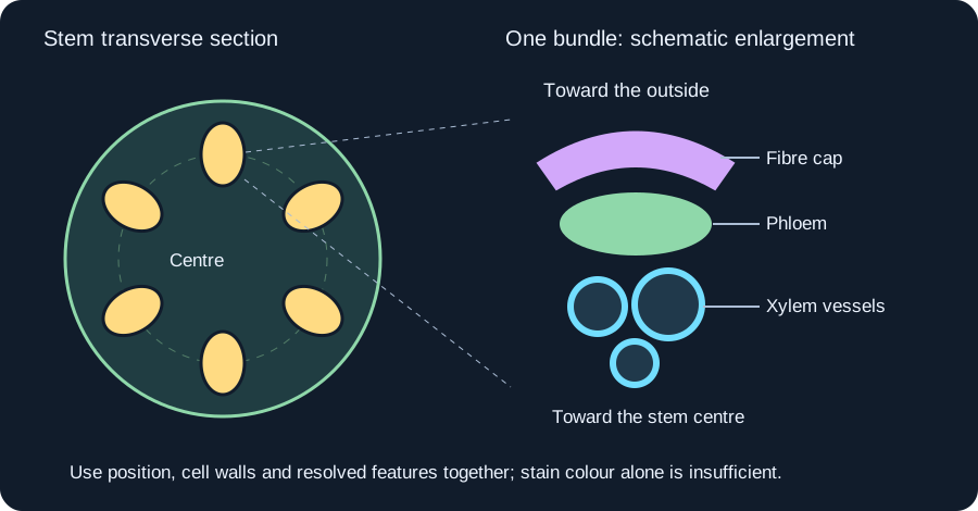
Locate the section at low power before using higher magnification and fine focus for detail. In a typical young dicot stem example, vascular bundles form a ring, with xylem toward the centre and phloem toward the outside. A sclerenchyma fibre cap may lie outside the phloem. This is an example arrangement, not a rule for every plant stem or developmental stage.
2
Identify tissue using several features
Xylem vessels commonly have a relatively large lumen and thick lignified walls, supporting water transport and resistance to collapse. Sclerenchyma fibres have thick lignified walls and support the organ. Phloem includes living transport-associated tissue; sieve tubes and companion cells have different features, some of which require an appropriate section and sufficient resolution to identify.
Use location, cell shape, wall appearance and an appropriate stain/reference together. A stain colour alone is not a unique tissue identity, and a large hole may be a preparation artefact. Do not draw a sieve plate or nucleus simply because a textbook says it exists if it was not resolved in the actual specimen.
3
Record observation before inferring function
Make a clear scientific drawing with representative proportions and labels that identify observed tissues. Add an appropriate scale bar or a measured magnification, keeping image and specimen units consistent. Record the plant material and section plane so another reader can interpret the arrangement.
Relate thick lignified walls to support or resistance to pressure change, and a lumen to a possible conducting pathway. A fixed section does not directly measure water flow, assimilate translocation or mechanical strength. Compare transverse and longitudinal information where useful and state uncertainty when preservation or resolution prevents a firm identification.
4
Worked applications
Attempt these problems on paper before opening the worked reasoning. Explain your steps as well as giving a final answer.
Example 1A vascular bundle is drawn 36.0 mm long on paper. Its corresponding measured specimen length is 0.900 mm. Find drawing magnification.
1Magnification = image length/actual length in matching units.
236.0/0.900 = 40.0.
Answer×40.0 for the stated drawing and measurement.
Example 2A region toward the inside of a dicot bundle has large lumens and thick lignified walls. What identity is supported, and what is not directly measured?
1The combined location and wall/lumen evidence supports xylem vessels in this context.
2The section does not directly measure the rate of water flow.
AnswerXylem is supported by the observed pattern; transport rate requires separate evidence.
Common mistakes to avoid
Identifying every tissue from stain colour alone.
Combine position, structure and suitable reference evidence.
Treating one dicot arrangement as universal.
Name the specimen and recognise developmental and plant-group differences.
Key takeaways
- You can prepare and orient a suitable stem section.
- You can identify tissue features and positions from observations.
- You can produce scaled drawings with justified structure–function links.
Apply it in an exam
Question 1: A region toward the inside of a dicot bundle has large lumens and thick lignified walls. What identity is supported, and what is not directly measured?
Show answer and reasoning
Xylem is supported by the observed pattern; transport rate requires separate evidence.
- The combined location and wall/lumen evidence supports xylem vessels in this context.
- The section does not directly measure the rate of water flow.
Explore further
- Edexcel 9BN0 official specification (opens in a new tab)
Check the assessed scope and practical or mathematical requirements.
- Read or explore this topic (opens in a new tab)
An external explanation or interactive activity to use alongside the lesson. It may include extension material.
Return to this tomorrow
Close the notes and answer from memory. Revisit any explanation you cannot yet reconstruct.
- Why use a thin section?
- What should a scientific drawing include?
Test yourself
Answer a few questions, then review anything you missed.
Next step
Browse practice topicsKeep practising
Start with the quiz above to check the key ideas. You can also choose another topic from the practice library.
Related lessons
All 18 lessons- Same chapterCP1: Caffeine and Daphnia Heart RateDesign a controlled invertebrate heart-rate comparison
- Same chapterCP2: Vitamin C and DCPIP ComparisonsStandardise a fixed DCPIP endpoint using a vitamin C reference
- Same chapterCP3: Membrane Permeability InvestigationsDesign a named-variable permeability investigation